

Enzyme-linked immunosorbent assay (ELISA) is widely used to detect and quantify antibodies in immunological research. However, selecting an appropriate assay kit becomes more complex when the target is a non-IgG antibody such as immunoglobulin A (IgA), immunoglobulin M (IgM), or immunoglobulin E (IgE). Unlike conventional IgG assays, these immunoglobulin classes have distinct structural characteristics, biological distributions, and analytical challenges that influence assay design and performance.
An effective non-IgG detection strategy requires more than selecting a kit with the correct antibody name. Researchers must consider whether the experiment measures total immunoglobulin concentration, antigen-specific antibody binding, or a particular molecular form. The selected ELISA format, capture reagents, detection antibodies, sample matrix, and calibration strategy can all affect the reliability of the results.
This article examines the major ELISA and binding assay formats used for IgA, IgM, and IgE detection, explains their technical advantages and limitations, and provides practical guidance for choosing suitable assay kits for different research objectives.

Why Do Non-IgG Antibodies Require Different Assay Strategies?
Although IgA, IgM, and IgE share the basic antigen-binding architecture of immunoglobulins, their structural and functional differences introduce important considerations for laboratory detection.
IgA is involved in mucosal immune responses and occurs in different molecular forms. Serum IgA is predominantly monomeric, while secretory IgA (sIgA) is commonly associated with polymeric IgA containing a joining chain and secretory component. These structural differences become particularly relevant when analyzing saliva, intestinal secretions, or other mucosal samples.
IgM is frequently produced during early stages of an adaptive immune response. Secreted IgM commonly forms pentameric structures, although hexameric forms can also occur. Its multivalent architecture supports avidity-driven binding but may complicate quantitative measurements and increase susceptibility to certain assay interferences.
IgE is typically present at much lower concentrations in circulation than IgG, IgA, or IgM. Its involvement in allergic sensitization and interactions with Fc epsilon receptors makes it an important target in immunology research. Detecting total IgE and measuring antigen-specific IgE require different analytical approaches.
Consequently, a successful non-IgG assay must account for the molecular form, expected abundance, and biological context of the target antibody. A format suitable for total serum IgA may not distinguish secretory IgA, while an indirect ELISA optimized for antigen-specific IgG may require substantial modification before reliable IgM or IgE measurements are possible.
Understanding the Main ELISA Formats for Non-IgG Detection
ELISA assays can be classified according to how the analyte is immobilized, captured, and detected. Direct, indirect, sandwich, and competitive approaches offer different benefits, depending on the target and experimental objective.
1. Direct and Indirect ELISA for Antibody Binding Detection
In an antigen-coated direct ELISA, an immobilized antigen captures specific antibodies from the test sample. A directly labeled antibody-specific detection reagent generates the signal.
In an indirect ELISA, an unlabeled sample antibody binds to the immobilized antigen, followed by detection with an enzyme-conjugated secondary antibody. This approach is commonly used to evaluate antigen-specific immune responses.
For non-IgG detection, the secondary antibody must recognize the appropriate immunoglobulin class. For example, an anti-human IgA antibody specific to the alpha heavy chain may be selected when measuring antigen-specific human IgA.
Indirect ELISA offers several practical advantages:
- Flexibility in choosing class-specific secondary antibodies.
- Signal amplification through enzyme-labeled detection reagents.
- Compatibility with multiple antigen targets.
- Suitability for comparing antigen-binding responses across experimental samples.
However, this format depends heavily on antigen immobilization quality and secondary antibody specificity. If the detection antibody cross-reacts with other immunoglobulin classes, the signal may not accurately represent the intended analyte.
Additionally, binding to an immobilized antigen does not necessarily reproduce interactions with the antigen in its native biological environment.
2. Sandwich ELISA for Total Immunoglobulin Quantification
Sandwich ELISA is commonly selected when the objective is to quantify an immunoglobulin in a biological sample.
In an antibody-capture sandwich format, a capture antibody is immobilized on the microplate surface. The target immunoglobulin binds to this reagent, and a second detection antibody recognizes a compatible, accessible epitope.
For example, a total IgA sandwich ELISA may employ an anti-IgA capture antibody and a noncompeting anti-IgA detection antibody.
This format can provide selective measurement of an immunoglobulin class in complex biological matrices. However, its performance depends on whether the capture and detection reagents recognize all relevant forms of the analyte.
A major consideration is epitope accessibility. Polymerization, molecular conformation, and interactions with other proteins can affect antibody recognition. Matched antibody pairs must therefore be evaluated for the immunoglobulin forms present in the intended samples.
3. Immunoglobulin-Capture ELISA for Antigen-Specific Antibodies
Class-capture ELISA is particularly useful when researchers need to detect antigen-specific antibodies while limiting interference from other immunoglobulin classes.
For instance, in an IgM-capture ELISA, immobilized anti-IgM antibodies first capture IgM from the sample. A labeled antigen, or an antigen followed by an appropriate detection system, is then used to identify the antigen-specific fraction.
This configuration differs from measuring total IgM because antigen recognition is an essential part of the detection process.
Similar concepts may be applied to IgA or IgE investigations, provided that suitable capture and antigen-detection reagents are available.
Class-capture formats can be advantageous when the target immunoglobulin is present alongside high concentrations of antibodies belonging to other classes. Nevertheless, antigen accessibility, capture efficiency, and competition between sample components still require careful evaluation.
4. Competitive ELISA for Alternative Quantitative Measurements
Competitive ELISA measures the ability of an analyte to compete with another binding component for a limited number of binding sites.
Depending on assay construction, sample antibodies may compete with labeled antibodies for antigen binding, or target immunoglobulins may compete with a labeled reference reagent.
In a conventional competitive assay, increasing analyte concentration often produces a decreasing signal. This inverse relationship must be considered during calibration and data interpretation.
Competitive ELISA can be useful when a suitable matched antibody pair is unavailable or when assay geometry makes a sandwich configuration unsuitable.
However, competitive formats are not automatically superior for complex non-IgG targets. Their suitability depends on reagent affinity, binding equilibria, assay dynamic range, and the specific molecular form being measured.
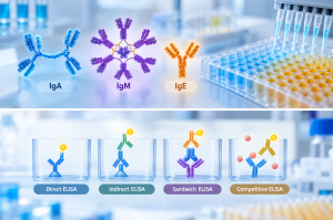
ELISA Format Comparison for IgA, IgM, and IgE Research
| Assay Format | Typical Research Objective | Main Consideration |
|---|---|---|
| Direct ELISA | Detect antibody binding to immobilized antigen | Requires appropriate labeled detection reagents |
| Indirect ELISA | Measure antigen-specific IgA, IgM, or IgE responses | Depends on secondary antibody class specificity |
| Sandwich ELISA | Quantify total IgA, IgM, or IgE | Requires compatible capture and detection antibodies |
| Class-capture ELISA | Detect antigen-specific antibodies within a selected isotype | Requires suitable antigen-based detection |
| Competitive ELISA | Quantify analytes through binding competition | Signal interpretation and calibration are format-dependent |
The optimal format is determined by the experimental question rather than the immunoglobulin class alone.
Choosing an IgA ELISA Kit: Total IgA, Secretory IgA, or Antigen-Specific IgA?
IgA detection is particularly important in mucosal immunology, vaccine research, and studies of local immune responses.
The first question when selecting an IgA assay kit is whether the objective is to quantify total IgA or identify a specific molecular form.
Total IgA Measurement
Total IgA ELISA kits are commonly used to measure IgA concentrations in serum, plasma, and other validated biological matrices.
A sandwich ELISA is often suitable for this purpose because class-specific capture and detection antibodies can recognize IgA independently of its antigen specificity.
However, not all total IgA assays respond equally to monomeric, dimeric, and polymeric IgA. Researchers should examine the kit’s documented analyte recognition and sample compatibility before assuming comparable recovery across different molecular forms.
Secretory IgA Detection
Secretory IgA is frequently studied in mucosal secretions, including saliva and intestinal samples.
A key analytical challenge is distinguishing sIgA from IgA lacking the secretory component.
An assay designed specifically for sIgA may combine recognition of the IgA alpha chain with detection of the associated secretory component. The complete assay architecture must be validated to distinguish the intended molecular complex from free secretory component or other sample constituents.
For mucosal research, selecting a general total IgA assay without confirming its specificity may lead to results that do not accurately represent the secretory IgA fraction.
Antigen-Specific IgA Analysis
When investigating mucosal responses to a defined antigen, antigen-coated indirect ELISA may be more appropriate than a total IgA assay.
The antigen captures antibodies with the relevant binding specificity, while an anti-IgA detection reagent identifies the IgA component of the response.
Antigen presentation is particularly important in this format. Denaturation during plate coating or poor surface orientation may alter accessible epitopes and influence the measured signal.
Researchers should therefore match the antigen preparation method to the biological interaction being investigated.
Choosing an IgM ELISA Kit: Addressing Multivalency and Interference
IgM presents distinct analytical challenges because of its large molecular size, polymeric organization, and multivalent antigen-binding properties.
Although IgM can interact strongly with multivalent antigen surfaces through avidity, this behavior should not be confused with high intrinsic affinity at each individual binding site.
Total IgM Quantification
For total IgM measurement, sandwich ELISA is commonly used with antibodies recognizing the IgM mu heavy chain.
The choice of capture and detection reagents is critical because both must retain access to their epitopes after IgM binding.
Polymeric structure and the potential presence of different IgM molecular forms can influence assay recovery. The calibration material should therefore be appropriate for the intended analyte, and dilution experiments should be performed when working with complex samples.
Antigen-Specific IgM Detection
Indirect ELISA and IgM-capture ELISA are both used to investigate antigen-specific IgM responses, but they differ in susceptibility to interference.
In an indirect assay, antigen is immobilized before adding the sample. Antigen-specific IgM binds to the surface and is detected using an anti-IgM reagent.
One limitation is competition from other antigen-specific immunoglobulins, especially IgG, which may occupy antigen-binding sites and reduce measurable IgM binding.
IgM-capture ELISA takes a different approach by capturing the IgM fraction before probing antigen specificity.
This configuration can help reduce some forms of immunoglobulin competition, although the actual improvement depends on assay design and validation.
Rheumatoid Factor and Nonspecific Binding
Rheumatoid factor, including IgM antibodies directed against IgG, can interfere with immunoassays containing IgG-based reagents.
Such interactions may contribute to false-positive signals in certain antigen-specific IgM assays. Conversely, competing antigen-specific IgG can produce falsely reduced IgM responses.
Possible mitigation strategies include sample pretreatment, validated blocking reagents, alternative capture configurations, and evaluation with appropriate interference controls.
These measures should not be applied indiscriminately because sample treatment may affect analyte recovery.
For IgM research, kit selection should prioritize demonstrated specificity and resistance to relevant interference rather than focusing exclusively on analytical sensitivity.
Choosing an IgE ELISA Kit: Sensitivity and Antigen Specificity
IgE detection requires particular attention to analytical sensitivity because circulating IgE concentrations are generally much lower than those of other major immunoglobulin classes.
The appropriate assay depends on whether the researcher intends to quantify total IgE or investigate binding to a defined antigen.
Total IgE Quantification
Total IgE sandwich ELISA typically uses anti-IgE antibodies for capture and detection.
Because IgE may be present at low concentrations, background control, antibody affinity, and signal detection chemistry are important considerations.
An assay with a low detection limit can be useful, but sensitivity alone is insufficient. Researchers should also evaluate the lower limit of quantification, precision near that limit, and the assay’s ability to distinguish low-concentration samples from background signals.
Antigen-Specific IgE Detection
Antigen-specific IgE assays are designed to identify IgE antibodies capable of binding particular allergens or other research antigens.
One common strategy immobilizes the antigen and detects bound IgE with an anti-IgE reagent.
An alternative reverse-capture strategy immobilizes anti-IgE antibodies before introducing a labeled antigen to identify antigen-reactive IgE.
These approaches have different analytical characteristics.
Antigen-coated formats may be influenced by competition from antigen-specific antibodies of other immunoglobulin classes. Reverse-capture methods can reduce certain forms of competition but introduce other constraints, including capture capacity and antigen-labeling requirements.
Researchers should assess how the selected format responds to samples containing different proportions of total and antigen-specific IgE.
Importantly, detection of antigen-specific IgE binding does not independently establish the biological activity of the antibody. Functional studies may require additional experimental systems that evaluate receptor engagement or cellular responses.
Key Factors to Consider When Selecting Non-IgG Assay Kits
A suitable assay kit should support the experimental objective, target antibody class, and sample conditions.
Several criteria deserve attention during selection.
1. Target Specificity and Species Reactivity
Confirm that the kit recognizes the intended immunoglobulin class and species.
For example, an anti-human IgM reagent should be assessed for cross-reactivity with human IgG, IgA, and IgE when these antibodies may be present in the sample.
Species reactivity is equally important when comparing human samples with animal models. Antibodies developed against one species may not recognize immunoglobulins from another species with equivalent affinity.
2. Detection Range and Sensitivity
The analytical measuring range should accommodate the expected target concentration after the required sample dilution.
A low detection limit does not necessarily mean that the assay can quantify the analyte accurately at that concentration.
Researchers should distinguish between the limit of detection and the validated quantitative range. Samples outside this range may require further dilution, a different assay, or additional validation.
3. Sample Matrix Compatibility
Serum, plasma, saliva, culture supernatants, and mucosal secretions contain different proteins and potential interfering substances.
Matrix components can alter antigen-antibody binding, affect enzyme reactions, or contribute to nonspecific background.
A kit validated with serum should not automatically be assumed suitable for saliva or tissue-derived fluids.
Matrix-specific recovery and dilution studies help determine whether the assay produces reliable measurements under the intended conditions.
4. Calibration Standards and Molecular Forms
Quantitative immunoassays depend on calibration materials.
For non-IgG antibodies, differences in polymerization, antigen-binding properties, and molecular composition may affect the relationship between the standard and the endogenous analyte.
Ideally, the calibration standard should represent the form of immunoglobulin being measured.
When this is not possible, researchers should recognize that assay-dependent concentrations may not be directly interchangeable between different kit formats.
5. Detection Chemistry and Available Equipment
Colorimetric ELISA remains widely used because it can be performed with conventional absorbance microplate readers.
Chemiluminescent immunoassays and other signal detection technologies may offer different sensitivity or dynamic range characteristics, depending on assay design.
However, improved signal generation does not eliminate problems caused by poor antibody specificity or sample interference.
Instrument compatibility, assay complexity, and reagent stability should all be considered alongside analytical performance.
How to Validate an ELISA Format Before Routine Use
Even a well-characterized commercial assay kit should be evaluated under the conditions of the intended study.
A practical qualification strategy includes several complementary experiments.
Standard Curve Evaluation: Prepare the required calibration series and confirm that the fitted curve supports reliable interpolation within the claimed working range. For many quantitative sandwich ELISAs, a four-parameter or five-parameter logistic model is appropriate when supported by the assay instructions.
Dilutional Linearity: Analyze samples across multiple dilution factors and assess whether dilution-corrected concentrations remain consistent. Unexpected changes can indicate matrix effects or other analytical problems.
Spike-and-Recovery Testing: Add known quantities of the target analyte to representative samples and determine how much can be recovered. This helps evaluate the influence of the sample matrix.
Parallelism Assessment: When suitable endogenous samples are available, compare their dilution responses with the calibration curve. This can reveal differences between the recognition of native analytes and reference standards.
Precision Testing: Assess variation among replicate wells and across independent assay runs. The required precision should be defined according to the study objective and assay performance expectations.
Specificity and Interference Controls: Include relevant negative controls, potential cross-reactants, and interference challenges when appropriate. These are particularly important for class-specific detection and IgM studies.
No single validation experiment demonstrates overall assay reliability. Confidence comes from combining specificity, recovery, precision, and matrix-compatibility evidence.
Common Problems in Non-IgG ELISA and How to Address Them
High Background Signal
High background may result from insufficient blocking, nonspecific secondary antibody binding, inadequate washing, or interference from sample components.
Optimization may involve adjusting blocking conditions, reagent concentrations, and washing procedures. Negative controls can help identify whether the problem originates from the sample or detection system.
Weak or Undetectable Signals
Low signals may reflect insufficient analyte concentration, inappropriate sample dilution, poor antibody affinity, or loss of antigen recognition after immobilization.
Before increasing signal amplification, researchers should confirm that the assay format is compatible with the target molecular form.
Inconsistent Results Across Dilutions
When calculated concentrations vary substantially with dilution, possible explanations include matrix interference, nonparallel calibration responses, or concentrations outside the assay’s quantitative range.
At very high analyte concentrations, some sandwich assays may also exhibit a high-dose hook effect, which can cause unexpectedly reduced signals.
Serial dilution studies can help identify these problems.
Different Results Between Commercial Kits
Two kits targeting the same immunoglobulin class may produce different measurements because they use different antibody pairs, standards, assay formats, or molecular recognition sites.
Such differences do not automatically indicate that either assay is defective.
For comparative studies, researchers should evaluate methodological consistency and avoid treating results from different assay systems as interchangeable without appropriate verification.
Frequently Asked Questions About Non-IgG ELISA Assay Kits
What Is the Difference Between Total IgA ELISA and Antigen-Specific IgA ELISA?
Total IgA ELISA measures the concentration of IgA regardless of antigen specificity. Antigen-specific IgA ELISA measures binding to a defined antigen. These assays answer different research questions and should not be used interchangeably.
Is IgM-Capture ELISA Always Better Than Indirect IgM ELISA?
Not necessarily. IgM-capture formats may reduce certain types of competition and interference, but their performance depends on capture efficiency, antigen presentation, and detection chemistry. Indirect IgM ELISA can also be suitable when properly optimized and validated.
Which ELISA Format Is Suitable for Low-Concentration IgE Detection?
A sensitive sandwich ELISA is often appropriate for total IgE quantification. For antigen-specific IgE, antigen-coated or reverse-capture formats may be considered. The selection should be guided by the expected concentration, antigen specificity, assay background, and available validation data.
Can the Same ELISA Kit Be Used for Serum and Saliva?
Only when the kit is validated or appropriately qualified for both matrices. Differences in protein composition, viscosity, analyte concentration, and potential interference may affect results. Salivary IgA research may also require specific attention to secretory IgA rather than total IgA.
Conclusion: Match the Assay Format to the Research Question
Selecting an assay kit for non-IgG antibody detection requires a clear understanding of the target immunoglobulin, its molecular characteristics, and the intended analytical readout.
For IgA research, distinguishing total, secretory, and antigen-specific measurements is essential. For IgM, polymeric structure, antibody competition, and assay interference must be considered. For IgE, sensitivity, background control, and the distinction between total and antigen-specific detection are particularly important.
Direct, indirect, sandwich, class-capture, and competitive ELISA formats each offer useful capabilities, but no single configuration is universally appropriate for all non-IgG antibody studies.
By aligning assay format selection with sample characteristics, analytical performance requirements, and experimental objectives, researchers can generate more interpretable and reproducible immunoglobulin detection data.
Related Non-IgG Antibody Products and Services from Creative Biolabs
Creative Biolabs provides research products and services supporting non-IgG antibody studies, including assay development, antibody discovery, and production-related research.
- Non-IgG Assay Kits — Browse immunoglobulin detection kits and compare available assay formats, species reactivity, and detection ranges.
- IgY-Based ELISA — Learn about the use of chicken-derived IgY antibodies as alternative recognition reagents in ELISA applications.
- Non-IgG Antibody Production and Purification — Explore production and purification solutions for non-IgG antibodies used in research and development.
- Non-IgG Therapeutic Antibody Discovery — Explore antibody discovery approaches for IgA, IgM, IgE, and other non-IgG antibody candidates.
For research projects involving non-IgG antibody detection or characterization, contact Creative Biolabs to discuss suitable products and research support options.
For Research Use Only. Not intended for diagnostic or therapeutic applications.