Systemic lupus erythematosus (SLE) remains one of the most heterogeneous and diagnostically challenging autoimmune diseases. Its complex pathogenesis involves dysregulated B cell responses and the persistent production of pathogenic autoantibodies, often reflected by elevated serum anti-dsDNA and anti-nucleosome IgG. However, the limited sensitivity and specificity of conventional blood-based biomarkers have led to growing interest in non-invasive, mucosal-derived alternatives—particularly those involving Immunoglobulin A (IgA), the most abundantly produced antibody isotype in the human body.
A recent study by Romero-Ramírez et al. (2023) sheds new light on the potential of salivary IgA subtypes (IgA1 and IgA2) as accessible, disease-relevant biomarkers for SLE. The work not only confirms elevated levels of these subclasses in patients with SLE but also reveals compelling correlations with clinical parameters, thus opening new possibilities for mucosal immunodiagnostics.
IgA and Its Underappreciated Role in Autoimmunity
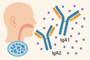
IgA accounts for nearly 70 mg/kg of daily antibody production and dominates mucosal secretions such as saliva, colostrum, and intestinal mucus. In humans, IgA exists in two subclasses:
-
IgA1, predominant in serum and saliva;
-
IgA2, enriched in colon and other secretions.
Both circulate largely as secretory IgA (SIgA) dimers in mucosal fluids, where they function as a first-line defense by neutralizing pathogens and modulating immune responses.
Despite its abundance, IgA’s clinical potential in autoimmune diagnostics has been underutilized. This is especially true for SLE, where diagnostic emphasis remains heavily skewed toward IgG-based autoantibodies, often missing mucosal contributions to systemic immune dysfunction.
Study Overview: Profiling Salivary IgA in SLE
To investigate this gap, Romero-Ramírez et al. designed a cross-sectional study involving 38 SLE patients (27 with inactive and 11 with active disease per SLEDAI criteria) and 14 age- and sex-matched healthy controls. The aim was to quantify salivary IgA1 and IgA2 levels and determine their relationship to established markers of lupus activity, such as anti-dsDNA titers and complement levels.
Saliva samples were collected through passive drooling, processed with protease inhibitors, and analyzed using a light chain capture ELISA for precise subclass quantification. Additionally, salivary autoantibodies—IgA anti-dsDNA and IgA anti-nucleosome—were measured via commercial ELISA kits adapted for IgA detection.
Core Evidence Supporting Salivary IgA as a Biomarker for SLE
1. Significant Elevation of IgA1 and IgA2 in SLE Saliva
The study revealed a clear increase in both IgA1 and IgA2 concentrations in the saliva of SLE patients compared to healthy individuals. IgA1 was the dominant subtype in all participants, but its levels were particularly elevated in active SLE patients, reaching values nearly five times higher than those in healthy controls.
Interestingly, total IgA (tIgA = IgA1 + IgA2) also demonstrated a strong association with disease activity, showing statistically significant elevation between inactive and active SLE groups.
2. Correlation with Serum Anti-dsDNA Antibody Titers
Through Spearman correlation analysis, positive associations were identified between salivary IgA subtypes and serum anti-dsDNA titers, one of the hallmark biomarkers of lupus nephritis and systemic flares. IgA1, IgA2, and tIgA all exhibited strong correlation coefficients, suggesting a systemic link between mucosal antibody production and systemic autoimmunity.
3. Discovery of Salivary IgA Anti-Nucleosome Antibodies
Perhaps the most novel finding was the detection of IgA-class anti-nucleosome antibodies in saliva, which were significantly elevated in SLE patients but virtually absent in healthy controls. This is particularly important given the established role of anti-nucleosome antibodies in SLE diagnosis and their ability to outperform anti-dsDNA in certain disease contexts.
Interestingly, no significant difference was found in salivary IgA anti-dsDNA levels between the two groups, possibly due to methodological limitations or mucosal compartmentalization of antibody responses.
4. Diagnostic Performance of IgA1 and IgA2
Receiver operating characteristic (ROC) curve analyses showed that salivary IgA1 achieved an AUC of 0.855, indicating outstanding discriminative capacity between SLE and healthy individuals. IgA2 also performed well (AUC = 0.761). These results are comparable to those of traditional blood-based biomarkers, emphasizing the clinical potential of salivary IgA subtyping as a non-invasive diagnostic adjunct.
Clinical Significance and Future Directions
The observed increases in IgA subtypes, particularly IgA1, raise intriguing possibilities about the mechanisms driving mucosal immunity in lupus. The dramatic rise in IgA1 levels could be linked to IL-10–mediated class switching, which is known to be dysregulated in SLE and elevated in both serum and saliva of affected individuals. Given that IgA1 has a long hinge region prone to proteolytic degradation, its persistence in saliva suggests active overproduction rather than passive leakage.
Moreover, the persistence of salivary IgA despite B cell depletion therapies like rituximab supports the idea that mucosal B cells or IgA+ plasmablasts may be more resistant to systemic immunosuppression—potentially maintaining a distinct immunological reservoir.
The use of saliva as a diagnostic biofluid also offers practical advantages: it is simple to collect, poses no infection risk, and allows for repeated, longitudinal monitoring—especially beneficial in pediatric or immunosuppressed populations.
Toward a New Paradigm in Lupus Biomarker Development
This study not only elevates the diagnostic potential of salivary IgA1 and IgA2 in SLE but also opens the door to broader translational research. For instance, tracking IgA glycoforms or Fc receptor interactions could yield insights into disease mechanisms or therapeutic targets. Moreover, the mucosal compartment’s role in systemic autoimmunity is likely underappreciated—and IgA is the key to unlocking it.
IgA-Related Services at Creative Biolabs
To support the growing research and clinical interest in IgA biology, Creative Biolabs offers a suite of custom services:
Whether you’re focused on mucosal immunology, autoimmune disease, or therapeutic antibody engineering, our platform is built to support your innovation.
Reference:
1. Romero-Ramírez, Sandra, et al. “Salivary IgA subtypes as novel disease biomarkers in systemic lupus erythematosus.” Frontiers in Immunology 14 (2023): 1080154.